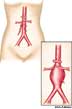
An abnormal, blood-filled sac formed by dilation of the wall of a blood vessel or heart ventricle, most commonly the abdominal aorta and intracranial arteries, resulting from disease or trauma to the wall, as in atherosclerosis.
[Middle English aneurisme, ultimately from Greek aneurusma, from aneurein, to dilate : ana-, throughout; see ana- + eurus, wide.]
(2-4) The pathogenesis of DA involves an arterial wall tear that leads to subintimal hematoma resulting in luminal narrowing, occlusion, aneurysmal dilatation, or vascular rupture.
Cardiac multidetector computed tomography showed coronary artery fistula (CAF) arising from the proximal left anterior descending artery with about 5-cm distal aneurysmal dilatation partially filled with thrombus.
Dynamic computed tomography scans showed fusiform aneurysmal enlargement in the proximal segment of the intrahepatic right portal vein (Figure 2a, 2b).
A fundus examination of the left eye revealed multiple telangiectatic and irregular, dilated aneurysmal retinal vessels, exudative retinal detachment with deep, and extensive subretinal lipid exudates with a well-defined macular edema.
These lesions include solitary bone cyst, aneurysmal bone cysts, osteofibrous dysplasia, myositis ossificans, brown tumour of hyperparathyroidism, and giant-cell granuloma.
More than half of these genes have previously been associated with pathological vascular phenotypes, and more than a third have specifically been associated with aneurysmal pathology.
All content on this website, including dictionary, thesaurus, literature, geography, and other reference data is for informational purposes only. This information should not be considered complete, up to date, and is not intended to be used in place of a visit, consultation, or advice of a legal, medical, or any other professional.