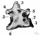
fo·ra·men
(fə-rā′mən)n. pl. fo·ram·i·na (-răm′ə-nə) or fo·ra·mens
An opening or orifice, as in a bone or in the covering of the ovule of a plant.
[Latin forāmen, an opening, from forāre, to bore.]
fo·ram′i·nal (-răm′ə-nəl), fo·ram′i·nous (-nəs) adj.
American Heritage® Dictionary of the English Language, Fifth Edition. Copyright © 2016 by Houghton Mifflin Harcourt Publishing Company. Published by Houghton Mifflin Harcourt Publishing Company. All rights reserved.
foramen
(fɒˈreɪmɛn)n, pl -ramina (-ˈræmɪnə) or -ramens
(Anatomy) a natural hole, esp one in a bone through which nerves and blood vessels pass
[C17: from Latin, from forāre to bore, pierce]
foraminal adj
Collins English Dictionary – Complete and Unabridged, 12th Edition 2014 © HarperCollins Publishers 1991, 1994, 1998, 2000, 2003, 2006, 2007, 2009, 2011, 2014
fo•ra•men
(fəˈreɪ mən)n., pl. -ra•mens, -ram•i•na (-ˈræm ə nə)
a small opening, orifice, or perforation, as in a bone or in the ovule of a plant.
[1665–75; < Latin forāmen hole, opening =forā(re) to bore1, pierce + -men resultative n. suffix]
fo•ram•i•nal (fəˈræm ə nl) adj.
Random House Kernerman Webster's College Dictionary, © 2010 K Dictionaries Ltd. Copyright 2005, 1997, 1991 by Random House, Inc. All rights reserved.
foramen
A hole in a bone or between two body cavities.
Dictionary of Unfamiliar Words by Diagram Group Copyright © 2008 by Diagram Visual Information Limited
ThesaurusAntonymsRelated WordsSynonymsLegend:
| Noun | 1. |  foramen - a natural opening or perforation through a bone or a membranous structure foramen - a natural opening or perforation through a bone or a membranous structureforamen of Monro, interventricular foramen, Monro's foramen - the small opening (on both the right and left sides) that connects the third ventricle in the diencephalon with the lateral ventricle in the cerebral hemisphere foramen magnum - the large opening at the base of the cranium through which the spinal cord passes |
Based on WordNet 3.0, Farlex clipart collection. © 2003-2012 Princeton University, Farlex Inc.